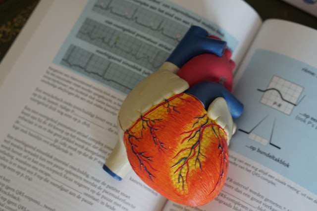

An electrocardiogram (ECG or EKG) is a test that measures the electrical activity of the heart, while an echocardiogram is a test that uses ultrasound to create images of the heart and its structures to evaluate its function and anatomy. In summary, ECG measures electrical activity while echocardiogram creates images using ultrasound.
What is an electrocardiogram?
(Image by OsloMetX from Pixabay )
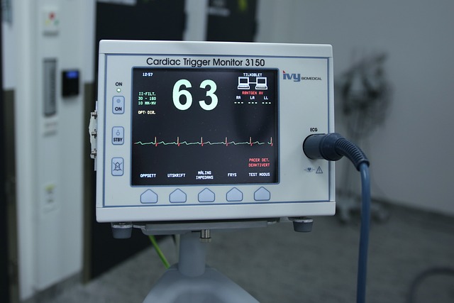
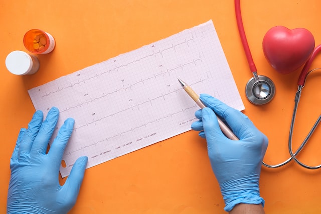
An electrocardiogram (ECG or EKG) is a medical test that records the electrical activity of the heart. It is a non-invasive and painless test that involves attaching electrodes to the chest, arms, and legs to detect the electrical impulses that occur as the heart beats.
During an ECG, the electrodes are connected to a machine that records the electrical activity and produces a graphical representation of the heart’s rhythm and activity. The test can help diagnose various heart conditions, including arrhythmias, heart attacks, and other abnormalities.
The ECG waveform consists of several waves that represent different parts of the cardiac cycle. The P wave represents the electrical activity that triggers the contraction of the atria, the QRS complex represents the electrical activity that triggers the contraction of the ventricles, and the T wave represents the recovery phase of the ventricles.
An ECG is a quick and non-invasive test that can be performed in a doctor’s office, hospital, or clinic. It is typically used as a routine part of a physical examination, to evaluate the heart’s function before surgery, or to monitor patients with known heart conditions.
What is an echocardiogram?
An echocardiogram is a non-invasive medical test that uses ultrasound waves to create images of the heart and its structures. It is a safe and painless test that provides detailed information about the heart’s size, shape, and function.
During an echocardiogram, a trained technician places a small, handheld device called a transducer on the chest or abdomen. The transducer sends high-frequency sound waves into the body, which bounce off the heart and create images on a computer screen.
Echocardiograms can provide valuable information about the heart’s function, including its ability to pump blood, the thickness and movement of its walls, and the functioning of its valves. The test can also detect abnormalities, such as blood clots, tumors, or fluid buildup around the heart.
There are several types of echocardiograms, including:
- Transthoracic Echocardiogram (TTE): This is the most common type of echocardiogram, which involves placing the transducer on the chest.
- Transesophageal Echocardiogram (TEE): This test involves inserting a small probe into the esophagus to get a closer view of the heart.
- Stress Echocardiogram: This test is performed while the patient is exercising on a treadmill or stationary bike to evaluate the heart’s response to exercise.
- Three-dimensional Echocardiogram: This test uses advanced imaging technology to create a 3D image of the heart.
Echocardiograms are used to diagnose and monitor a variety of heart conditions, including heart valve disease, congenital heart defects, heart failure, and other heart abnormalities. The test is typically performed in a hospital or clinic and takes about 30-60 minutes to complete.
The Difference between the Two
The main difference between an electrocardiogram (ECG) and an echocardiogram (ECHO) is that an ECG measures the electrical activity of the heart while an ECHO image shows the structure and motion of the heart.
An electrocardiogram is a test that records the electrical activity of your heart through sensors called electrodes that are placed on your skin. This test can show if there are any problems with your heart’s rhythm or if there is evidence of a previous heart attack.
An echocardiogram uses ultrasound waves to create a moving image of your heart. This test can show how well your heart is pumping, if there is any damage to your heart, and if there are any abnormal growths.
When is an Electrocardiogram needed?
An electrocardiogram (ECG) is a test that measures the electrical activity of your heart. An ECG shows how fast your heart is beating and the strength and timing of your heartbeats.
Your doctor may recommend an ECG if you have symptoms of heart problems, such as chest pain or shortness of breath. An ECG also may be done to check for signs of heart disease, such as a previous heart attack, and to monitor the health of your heart after a heart attack or other treatment.
When is an Echocardiogram needed?
An echocardiogram is a type of ultrasound test that uses sound waves to create moving images of your heart. The test is also called an echo.
Your doctor may recommend an echocardiogram to check for problems with the structure and function of your heart. An echocardiogram can be used to:
- Detect heart disease
- Determine the cause of unexplained chest pain
- Evaluate the effectiveness of treatments for a heart condition
- Monitor the progress of known heart conditions, such as congestive heart failure
- Detect abnormal heart rhythms (arrhythmias)
An echocardiogram is generally a safe procedure with little or no discomfort. You may feel slight pressure on your chest from the transducer, but this should not be painful.
How to Prepare for an Electrocardiogram
Preparing for an ECG is easy. No special preparation is needed, and you can eat and drink normally before the test. You will need to remove any clothing or jewelry that might interfere with the test. You will also need to remove any lotion, oils, or makeup from your skin so that the electrodes can make good contact.
How to Prepare for an Echocardiogram?
If you’re scheduled for an echocardiogram, you may be wondering how to prepare. Here are a few things you can do to make sure your test goes smoothly:
- First, check with your insurance provider to make sure the test is covered.
- Next, schedule the test for a time when you can relax and won’t be rushed.
- On the day of the test, wear comfortable clothing that doesn’t have metal snaps or zippers.
- You’ll also need to remove any jewelry, including body piercings.
- Finally, it’s important to fast for four hours before your echocardiogram. This means no food or drinks, not even water.
Is ECG same as echocardiogram?
No, an ECG (electrocardiogram) is not the same as an echocardiogram. While both tests provide important information about the heart, they use different methods to do so.
An ECG measures the electrical activity of the heart and provides information about the heart’s rhythm and rate. It is a non-invasive test that involves attaching electrodes to the chest, arms, and legs to record the electrical activity of the heart.
On the other hand, an echocardiogram uses ultrasound waves to create images of the heart and its structures. It provides information about the heart’s size, shape, and function, including the thickness and movement of the heart’s walls and the functioning of its valves. It is also a non-invasive test, but it involves placing a transducer on the chest or abdomen to create images of the heart.
Both tests are important diagnostic tools used to evaluate heart function and detect abnormalities. They are often used in combination with other tests and examinations to provide a complete picture of the heart’s health.
What is the difference between electrocardiogram and electrocardiograph?
An electrocardiogram (ECG) is a test that measures the electrical activity of your heart. An electrocardiograph (EKG) machine records these signals on a paper or computer.
The main difference between an ECG and an EKG is that an ECG measures the electrical activity of your heart, while an EKG machine records these signals on a paper or computer.
An ECG can help diagnose conditions such as heart attacks, irregular heart rhythms, and cardiomyopathy. An EKG may also be used to monitor the effects of certain medications or procedures on your heart.
What are the two types of echocardiogram?
An echocardiogram is a type of ultrasound test that uses sound waves to create images of your heart. The test is also called an echo. An echocardiogram can help your doctor evaluate the size and shape of your heart and how well your heart is functioning. There are two types of echocardiograms:
Transthoracic echocardiogram: This is the most common type of echocardiogram. The test is performed with a hand-held device called a transducer, which is placed on your chest. The transducer sends out sound waves that bounce off your heart and create echoes. These echoes are converted into images that are displayed on a monitor.
Transesophageal echocardiogram: This type of echocardiogram is usually done if a transthoracic echocardiogram does not provide enough information about your heart or if you have certain heart conditions, such as atrial fibrillation. In this test, the transducer is passed down your throat and into your esophagus (the tube that connects your mouth to your stomach). The transducer gets closer to your heart than in a transthoracic echocardiogram, so it can provide more detailed information about your heart.
Can echo detect blockage?
An echocardiogram (echo) may be able to detect certain types of blockages in the heart, depending on the location and severity of the blockage.
An echocardiogram can detect blockages in the heart’s blood vessels, such as the coronary arteries, by evaluating the heart’s function and blood flow. If the heart muscle is not receiving enough blood due to a blockage, the echocardiogram may show areas of the heart that are not functioning properly or that have reduced blood flow. This may be seen as abnormal wall motion or reduced ejection fraction, which is the amount of blood pumped out of the heart with each beat.
However, an echocardiogram may not be able to detect all blockages, particularly those that are small or located in certain areas of the heart. In these cases, other tests, such as a cardiac catheterization or a coronary angiogram, may be necessary to accurately diagnose the blockage.
It’s important to note that an echocardiogram is a non-invasive and safe test that can provide valuable information about the heart’s function and structure. If your doctor suspects that you have a blockage in your heart, they will likely use a combination of tests and examinations to make a diagnosis and determine the best course of treatment.
Featured Image By – Photo by Robina Weermeijer on Unsplash